Beef Marbling Chart Ultrasound Scan Data
- History
- Principles of Carcass Ultrasound
- Measurements Collected
- Data Collection Procedure
- Using Carcass Ultrasound Data
- Literature Cited
The beef industry has begun utilizing a value-based marketing system, where cattlemen are rewarded for producing a high quality, consistent end product that meets consumer demands. One tool that aids producers in the efficient and profitable production of beef cattle is live animal carcass ultrasound. Carcass ultrasound is an economical way cattlemen can make genetic improvements in carcass traits, which will, in turn, put profits in their pockets.
History
The use of ultrasound was first reported in the 1950s and the technique has been utilized in the beef industry for more than 40 years (Houghton and Turlington, 1992). Before the development of live animal carcass ultrasound, the only way to determine the carcass merit of a sire was to evaluate its slaughter progeny through the feedlot and packing plant. Carcass traits were measured in the progeny and the data were analyzed through a genetic evaluation program to produce carcass trait EPDs (Expected Progeny Differences). This process was slow, laborious and expensive. The average time period to "prove" a sire for carcass merit was four to seven years, at a cost of $5,000 to $10,000. Because of these constraints, relatively few bulls had enough data to calculate accurate and meaningful carcass EPDs. Using ultrasound technology, progeny testing for carcass traits can be completed in as short as two years and for as little as $1,000 per sire evaluated (Williams, 2001). Because live animal ultrasound is non-invasive, carcass data can be collected on a much larger population of animals, including yearling bulls and heifers from seedstock producers and half-sib steers and heifers from commercial producers.
Principles of Carcass Ultrasound
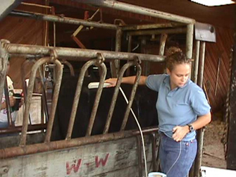
The ultrasound technology used for carcass trait measurement is referred to as real-time ultrasound. Real-time ultrasound uses high frequency sound waves (generally 2 to 10 MHz) to "see" under the animal?s hide while it is still alive. This is the same technology used for pregnancy diagnosis in both livestock and humans. A sound-emitting probe, or transducer, is placed snuggly on the animal?s back and the sound waves penetrate the tissues, reflecting off the boundaries between hide, fat and muscle layers. As the sound waves reflect back towards the probe, a cross-sectional image is created on the ultrasound machine monitor, which allows measurement of the various carcass traits. This process is harmless to the animal and the technician (Houghton and Turlington, 1992).
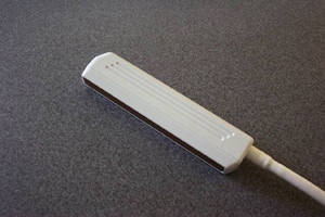 Figure 1. An ultrasound transducer houses tiny crystals that emit and catch the sound waves.
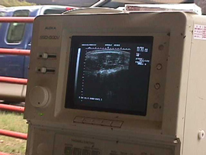 Figure 2. The ultrasound machine displays the reflected sound waves as an image on the screen. This is a ribeye image.
Measurements Collected
The measurements collected utilizing live animal carcass ultrasound can be used to estimate carcass retail yield and meat quality. The common traits estimated include ribeye area (REA), backfat (BF), rump fat (RF) and percent intramuscular fat (PIMF). Ribeye area, in square inches, is measured between the 12th and 13th ribs and gives an estimate of the amount of muscle and lean product in the animal. Backfat, in inches, is also measured between the 12th and 13th ribs and is an estimate of the external fat on the animal. This measurement is taken at a point three-fourths of the length of the ribeye from the end nearest the animal?s spine and is the most important factor affecting retail product yield. Rump fat is an additional measure of external fat on the animal and is also measured in inches. This measurement is taken along the rump of the animal between the hooks and pins. Percent intramuscular fat is an objective measurement of marbling in live cattle. Marbling is the main trait used to determine USDA Quality grades, thus PIMF gives a good indication of the animal?s meat quality (Perkins et al., 2003).
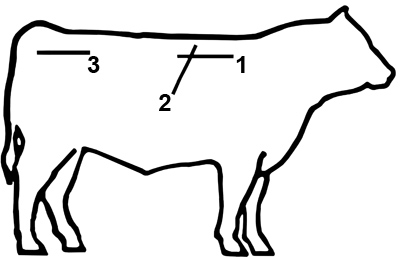 Figure 3. Ultrasound measurements are collected at three points on the animal (Silcox, 2002).
1. Percent intramuscular fat
2. Ribeye area and backfat
3. Rump fat
Data Collection Procedure
After carefully considering the advantages of using real-time, live animal carcass ultrasound, the producer is ready to take the next step and begin the data collection process. There are a variety of things to consider before jumping in, including proper preparation and adherence to the guidelines adopted by the corresponding breed association to ensure image collection and interpretation go smoothly.
Young seedstock animals (bulls, steers and developing heifers) can be used to collect ultrasound measurements. In order for accurate genetic differences to be expressed, the animals should be growing at a rate that will allow differences in lean and fat deposition to be measured. The ideal time to scan seedstock cattle is at 365 days of age. Due to the variation in calving dates within a herd, scanning age windows have been developed such that the data can be adjusted to yearling age. These windows allow a whole contemporary group of animals to be scanned together. The development of ultrasound EPDs requires that scanned animals be included in a well-defined contemporary group. A contemporary group includes all animals of the same breed and sex born in a given calving season, and reared and managed under similar conditions. These age windows vary among cattle breeds. The age windows for breeds may change periodically based on new research findings, so it is important to contact the breed association to determine the most current age requirements.
| Table 1. Some of the age windows for various breeds of cattle. | |||
| Breed | Yearling Bulls | Developing Heifers | Feedlot Steers & Heifers (days) |
| Angus | 320-440 | 320-460 | 320-480 |
| Brangus | 310-600 | 310-600 | 310-600 |
| Charolais | 320-430 | 320-430 | 320-430 |
| Hereford | 301-530 | 301-530 | 301-530 |
| Simmental | 270-500 | 270-500 | 270-500 |
| Gelbvieh | 320-420 | 320-420 | 320-420 |
After determining the correct scanning time for a group of cattle, the producer needs to contact a "certified" ultrasound technician. In order for their data to be accepted by the various breed associations, ultrasound technicians are required to meet a set of minimum standards for image quality, data accuracy and knowledge of ultrasound technology determined by the Ultrasound Guidelines Council (UGC). Only images collected by a UGC "certified" technician can be used to calculate carcass ultrasound EPDs. A list of certified technicians can be obtained from the breed association and in many cases will be published in breed association journals. Shopping around for a certified technician is a good idea. A local technician is the most obvious initial choice, simply because of location and ease of scheduling. Scheduling with a technician may be easier if scanning is coordinated with other breeders in your area, particularly if there are a relatively small number of cattle to scan (< 30 head). With more cattle to scan, technician travel and set expenses should decrease. Simply schedule ultrasound data collection with neighbors to reduce costs or gather cattle at a central location. Most technicians charge a flat fee per animal plus interpretation and travel expenses; therefore, technicians who are closer will probably have lower travel expenses. Most technicians charge between $15 and $25 per animal, with variable rates for larger numbers of cattle measured.
It is the producer?s responsibility to obtain all necessary paperwork and have the facilities in proper working order. The producer should contact the breed association and inform them of his/her intentions to collect ultrasound data. The association will then send barnsheets that include all necessary information, such as: registration numbers, date of birth, sex, etc?. The producer will need to fill in management codes and contemporary group codes. The breed association can help with these forms and describe their specific requirements. It is also the producer?s responsibility to ensure that their cattle-handling facilities are adequate for animal restraint and for the safety of both the technician and the cattle. A squeeze chute with fold-down side panels is best. A clean working area with a grounded power source (110v) is also required, preferably chute-side. It is best if there is a dedicated circuit to the chute-side power source, free from electrical interference that may be caused by other equipment, such as electrical motors. The producer should discuss the facilities with the technician so that proper arrangements can be made (Silcox, 2002).
On scan day, the ultrasound technician will save the necessary images for EPD calculation and send them along with the completed barnsheet to a centralized processing lab for interpretation. The general process for image collection begins with clipping the animal?s hair to within ½ inch to improve image quality in the 12th ? 13th rib region and over the rump. Next, a minimum of six images are collected: a cross-sectional image between the 12th and 13th ribs for measuring backfat and ribeye area; an image collected between the hooks and pins for determining rump fat; and four images taken parallel to the backbone and between the 12th and 13th ribs for estimation of marbling. Additional images may be collected to ensure optimum image quality. It is also necessary to weigh the cattle being scanned within seven days of the scan date (Perkins et al., 2003). Weights should be taken in the morning prior to feeding. It is preferred, although not required, to hold animals off both feed and water overnight. This weight is the prediction of empty body weight and thus gut fill should be minimized.
The images must be interpreted by a centralized lab for use in EPD calculation. The lab will then send the interpreted measurements to the respective breed association, who will use them in their genetic evaluation to develop carcass trait EPDs. The data will then be sent from the breed association to the breeder for his/her use. Turnaround time generally takes 14 days from the time of image collection.
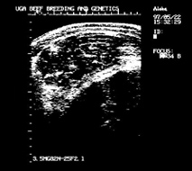 Figure 4. Ultrasound image of ribeye taken between 12th and 13th ribs.
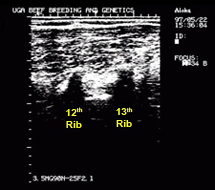 Figure 5. Longitudinal image used to measure percent intramuscular fat.
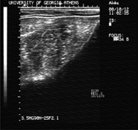 Figure 6. Rump fat image taken between the hooks and pins.
Using Carcass Ultrasound Data
Ultrasound carcass measurements are no different than traits like birth, weaning and yearling weight in that environmental conditions (nutrition and weather) play a role in expression of these traits. Therefore, it is more desirable to make selection decisions based on relative differences within a contemporary group (carcass trait ratios) rather than the actual measurements. Better still is the use of ultrasound carcass EPDs, if they are available for the breed. Since ultrasound carcass traits are generally considered highly heritable, selection of bulls and heifers or cows based on these traits will result in genetic change in their calves.
As with other traits of economic importance, producers should have a plan for utilizing ultrasound carcass information in the herd. This ensures that the producer will make focused and educated selection decisions that best fit the particular program rather than chasing the latest fads. Two key components of this plan are: 1) a "snapshot" of the herd?s current performance level, with respect to carcass value and 2) a defined "target" of the types of cattle, and, subsequently, types of carcasses, that can be produced while maximizing profits. The difference between the "snapshot" and the "target" will define the selection program and the breeding stock that will be selected.
There are a variety of ways to develop the "snapshot." Participation in a retained ownership program or some other value discovery program that provides feedback on the carcass value of feeder cattle is probably the best approach. An alternative would be simply to collect ultrasound data on the yearling cattle in the herd and utilize those measurements as the "snapshot"; however, this may not be economically feasible for some commercial producers. Identifying the "target" may not be as easy as it sounds either. The "target" is how and where a producer markets his cattle. Whether a producer is marketing seedstock (bulls to commercial cattlemen or replacement heifers) or feeder cattle for a terminal market makes a difference in how he/she will utilize the ultrasound data. Once the "snapshot" and "target" are identified, ultrasound carcass EPDs are simply the tools used to make genetic progress toward the goal of a profitable product.
Breeders should keep "common sense" in mind when developing a selection program that includes carcass traits. Ultrasound carcass EPDs can be used to make genetic changes, but concentrating solely on carcass traits while ignoring other production traits, especially reproductive traits, will certainly lead to trouble. For instance, continued selection against backfat will almost certainly result in reduced reproductive performance of the cowherd. Also, ribeye area and mature size are genetically related so that selection for ribeye area will generally result in increased frame size and ultimately could negatively impact reproductive performance. Fortunately, there does not appear to be any significant downside for the cowherd in response to selection for increased marbling. But, producers should never utilize single trait selection (Wall, 2005).
Real-time live animal carcass ultrasound can be a beneficial production practice for all segments of the beef industry. This technology is being utilized across the nation by progressive purebred and commercial bull producers and buyers as they integrate more carcass information into their selection programs. The use of live-animal carcass ultrasound is just one step towards reaching the goal of producing a high-quality, consistent product for today?s value-based market.
Literature Cited
Houghton, P.L., and L.M. Turlington. 1992. Application of ultrasound for feeding and finishing animals: A review. J. Anim. Sci. 70: 930-941.
Perkins, T., A. Meadows, and B. Hays. 2003. Study Guide for the Ultrasonic Evaluation of Beef Cows for Carcass Merit. Ultrasound Guidelines Council.
Silcox, R. Editor. 2005. Guidelines for the Uniform Beef Improvement Programs. Beef Improvement Federation. 8: 37-44.
Wall, P. 2005. Understanding the Ultrasound Info Craze. www.cuplab.com/location.cfm? fuseaction=dspThisArticle&newsId=38
Williams, A.R. 2001. Live Animal Carcass Ultrasound, Can it Benefit You? Mississippi State University. P2253. 1-2.
Status and Revision History
Published on Feb 05, 2008
Published on Feb 04, 2009
Published on Feb 28, 2011
Published with Full Review on Feb 01, 2014
Published with Minor Revisions on Jan 01, 2018
Source: https://extension.uga.edu/publications/detail.html?number=B1337&title=Using+Live+Animal+Carcass+Ultrasound+in+Beef+Cattle
0 Response to "Beef Marbling Chart Ultrasound Scan Data"
Post a Comment